Retinal imaging eye test could detect early Frontotemporal dementia
Scientists think they have discovered a simple way of diagnosing Frontotemporal dementia, through a retinal imaging test.
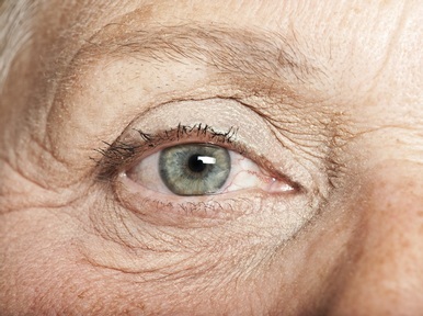
Researchers from the Perelman School of Medicine at the University of Pennsylvania carried out a study using a non-expensive and non-invasive eye-imaging technique and found that people with Frontotemporal dementia (FTD) showed a thinning of the outer retina compared to people without the disease.
Lead author of the study, Benjamin Kim, assistant professor of Ophthalmology at Penn’s Scheie Eye Institute said: “Our finding of outer retina thinning in this carefully designed study suggests that specific brain pathologies may be mirrored by specific retinal abnormalities.”
FTD which is a rare type of dementia, is caused by damage to the brain cells in the frontal lobe. These cells regulate behaviour and emotions as well as the temporal lobe which enables the understanding and production of language.
It is hoped that earlier diagnosis of the disease will result in more effective treatment.
The study’s senior author, Murray Grossman, a professor of Neurology and director of the Penn FTD Center, said: “As we enter an era of disease-modifying treatments for neurodegenerative disorders, it is essential for us to have tools that can identify the specific pathologies accumulating in the brain so that we can administer the appropriate treatments to patients who are likely to benefit.”
The study, which was published in Neurology, found the thinning of retinas of the people with FTD was 10 per cent more than the people without the disease. In addition it found thinning worsened in the people whose cogitation test results were lower.
The Penn researchers now plan to carry out larger, more conclusive studies to compare retinal measurements among patients who have different FTD subtypes as well as other neurodegenerative diseases.
Latest News
 29-Jul-24
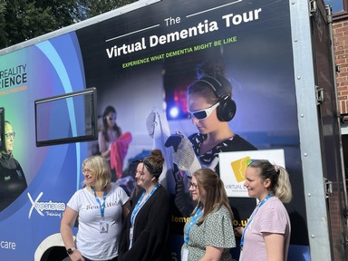
Dementia Bus gives carehome.co.uk staff insight into life with dementia
29-Jul-24
Dementia Bus gives carehome.co.uk staff insight into life with dementia
 01-Mar-24
Find out the top care homes in 2024
01-Mar-24
Find out the top care homes in 2024
 21-Mar-23
UK's top care homes in 2023 revealed
21-Mar-23
UK's top care homes in 2023 revealed
 03-Jan-23
carehome.co.uk launches free care helpline
03-Jan-23
carehome.co.uk launches free care helpline
 13-Dec-22
5 mins with Emily Whitehurst, chief operating officer for Constantia Healthcare
13-Dec-22
5 mins with Emily Whitehurst, chief operating officer for Constantia Healthcare